Case Study
The patient is a 50 year-old female. She is edentulous in the upper left maxillary lateral incisor region (no. 10). For esthetic as well as functional reasons, she wants that area reconstructed. A CBCT scan was taken. The treatment plan was an implant and cemented crown. Subsequently, a 3.5 x 10mm Zuga implant with a sealing cap was placed. No temporary was used as the patient had that edentulous areas for several years. The medical history was essentially negative and non-contributory (Fig. 1-8).
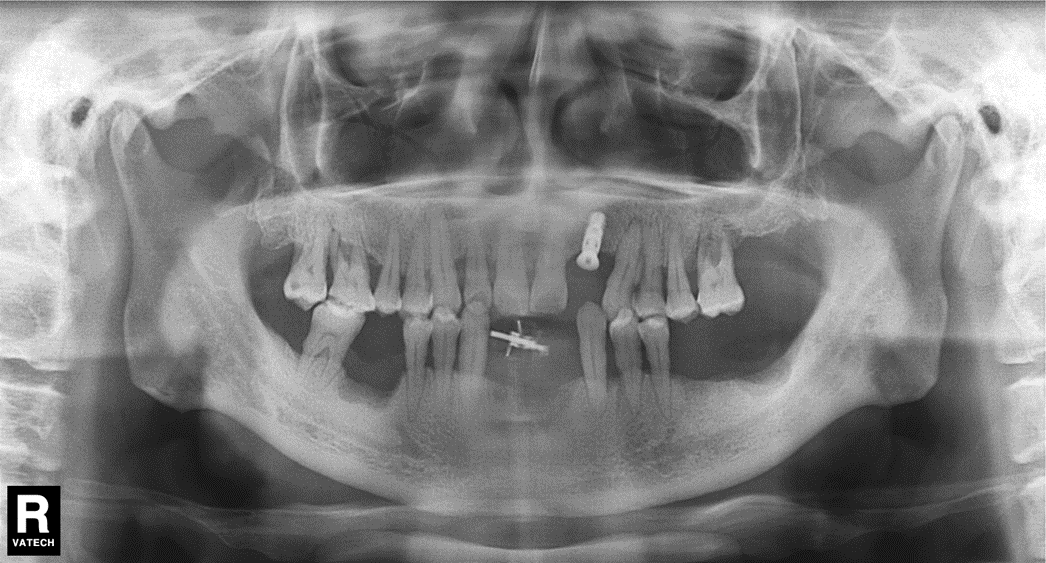
Figure 1. A panoramic radiograph demonstrating the edentulous area at left maxillary lateral incisor.
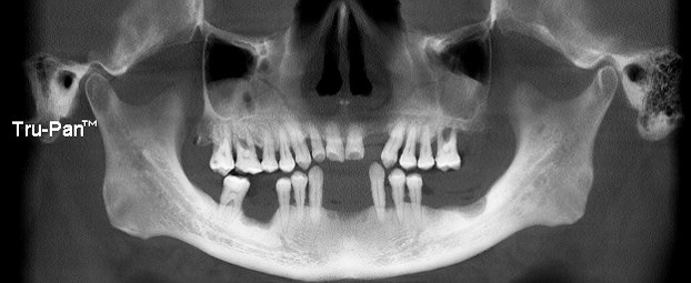
Figure 2a. The clinical view of the edentulous area with the mucoperiosteal flaps reflected.
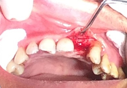
Figure 2b. The clinical view of the 3.5 x 10mm Zuga implant in place.
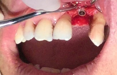
Figure 2c-2d. The clinical view with the implant and sealing cap in position.
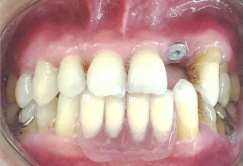
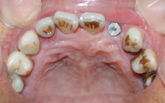
Figure 3. A panoramic radiograph with the 3.5 x 10mm Zuga implant and sealing cap in place.